Vasculearn Network · Clinical Hub
Atherosclerotic Cardiovascular Disease (ASCVD)
Printed November 14, 2026 · vln.org/hcp/disease-state/ascvd
Clinical Overview
ASCVD includes coronary, cerebrovascular, and peripheral artery disease (PAD) caused by atherosclerosis. This page focuses on coronary ASCVD and lipid management, from identifying risk before a first event to reducing the risk of recurrence.
Atherosclerosis develops as cholesterol-containing particles accumulate in artery walls and inflammation contributes to plaque growth. Plaque can limit blood flow, while plaque disruption can trigger thrombosis and an acute coronary event. Imaging may identify subclinical disease before symptoms develop.
Established clinical ASCVD and subclinical atherosclerosis call for different approaches to risk assessment, treatment intensity, and lipid goals.
Coronary artery disease
Atherosclerotic coronary artery disease (CAD) may present with angina or exertional dyspnea or remain asymptomatic. Plaque disruption and thrombosis can cause acute coronary syndrome (ACS), including unstable angina and myocardial infarction (MI).
Plaque detected on imaging shouldn’t automatically be treated as equivalent to prior MI or coronary revascularization. Its extent and the patient’s overall risk help determine the appropriate preventive treatment.
Cholesterol and cardiovascular risk
LDL cholesterol (LDL-C) is a major treatment target, but it doesn’t tell the whole risk story. Non–HDL cholesterol reflects cholesterol carried by all atherogenic particles. Apolipoprotein B (ApoB) and lipoprotein(a), or Lp(a), can help clarify risk beyond LDL-C.
Treatment intensity depends on the patient’s clinical history, overall risk, and the LDL-C reduction needed to reach their goal.
Diagnosis
Assess lipids and inherited risk
Lipid panel
Most patients can have a nonfasting lipid panel for routine screening and risk assessment. Obtain a fasting panel when the patient has:
- Nonfasting triglycerides ≥400 mg/dL
- A known or suspected triglyceride metabolism disorder
- A family history of dyslipidemia or premature ASCVD
Fasting improves triglyceride assessment and can help estimate LDL-C when nonfasting triglycerides are high. Evaluate secondary causes of abnormal lipids and consider familial hypercholesterolemia (FH) when LDL-C is markedly elevated.
Lp(a)
Measure Lp(a) at least once in adulthood, and note that it isn’t included in a standard lipid panel. Levels ≥125 nmol/L or ≥50 mg/dL indicate increased risk. These are separate unit-specific thresholds, not a fixed conversion. Use the result to refine risk discussions, address other risk factors, and guide family screening.
ApoB
Consider ApoB when triglycerides are elevated (particularly ≥150 mg/dL), diabetes is present, or achieved LDL-C is low (<70 mg/dL). It can identify residual atherogenic particle burden even after LDL-C and non–HDL-C goals are met.
Estimate risk before a first event
Use PREVENT-ASCVD for eligible adults aged 30–79 years without clinical ASCVD or subclinical atherosclerosis and with LDL-C of 70–189 mg/dL. In eligible adults aged 30–59 years, consider 30-year risk as well. Discuss risk enhancers that the calculator doesn’t capture.
Personalize risk assessment with risk enhancers
The PREVENT-ASCVD estimate informs the discussion but risk enhancers can help personalize it. Examples include family history of premature ASCVD, chronic inflammatory conditions, higher risk ancestry, and reproductive history, including early menopause, preeclampsia, gestational diabetes, and preterm delivery. These may influence treatment decisions, particularly when estimated risk is borderline or intermediate.
Use coronary testing when it can change care
Coronary artery calcium
Consider coronary artery calcium (CAC) scoring in men aged ≥40 years and women aged ≥45 years when a primary prevention treatment decision remains uncertain, particularly at borderline or intermediate risk.
- A score of zero doesn’t exclude noncalcified plaque or override every other indication for treatment.
- CAC scoring isn’t a substitute for evaluating chest pain.
Symptoms suggesting coronary disease
New or worsening chest discomfort or other symptoms concerning for ACS require urgent assessment. For stable symptoms, select testing through a chest pain evaluation pathway based on clinical likelihood, prior disease, and patient characteristics.
Treatment
Secondary prevention: Lower LDL-C in established ASCVD
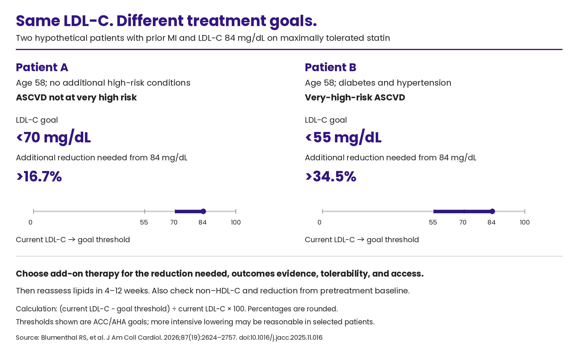
Use a high-intensity or maximally tolerated statin, adding nonstatin therapy when needed. Assess both the percentage reduction and the level reached.
The 2026 ACC/AHA multisociety dyslipidemia guideline sets lipid goals for established clinical ASCVD based on whether the patient meets very-high-risk criteria. Use these goals to assess response to maximally tolerated statin therapy and the need for additional lipid lowering.
Clinical ASCVD category | LDL-C goal | Non–HDL-C goal |
|---|---|---|
Not at very high risk | <70 mg/dL | <100 mg/dL |
Very high risk | <55 mg/dL | <85 mg/dL |
Who qualifies as “very high risk”?
Patients with multiple major ASCVD events, or one major event plus multiple high-risk conditions.
Major ASCVD events include:
- ACS within the past 12 months
- Prior MI, excluding the recent ACS already counted
- Ischemic stroke
- Symptomatic PAD
High-risk conditions include:
- Age >65 years
- Prior coronary bypass surgery or percutaneous coronary intervention (PCI) outside the major ASCVD event already counted
- Current smoking
- Diabetes
- Hypertension
- History of congestive heart failure
- LDL-C >100 mg/dL despite maximally tolerated statin therapy plus ezetimibe
Example: a patient with a prior MI, diabetes, and hypertension qualifies as very high risk. Coronary revascularization alone is a high-risk condition, rather than a major event in this classification.
When to add nonstatin therapy
In established ASCVD, add therapy when maximally tolerated statin treatment doesn’t achieve the patient’s lipid goals, generally LDL-C <70 mg/dL, or <55 mg/dL for very-high-risk patients. Confirm adherence and assess the additional reduction needed.
- Very-high-risk ASCVD: Add ezetimibe and/or a PCSK9 monoclonal antibody (evolocumab or alirocumab) to reach goal. Bempedoic acid is another guideline-supported option when additional lowering is needed.
- ASCVD not at very high risk: Ezetimibe, a PCSK9 monoclonal antibody, or bempedoic acid are reasonable options. Choose based on the reduction needed, patient preference, tolerability, and access.
There’s no requirement to try every agent in order. Some patients need combination therapy to reach goal.
Where inclisiran fits
Check response and tolerability
- Evaluate reported statin symptoms, interactions, and other possible causes; adjust treatment rather than leaving LDL-C unaddressed.
- Repeat a lipid panel 4–12 weeks after starting or adjusting therapy, then every 6–12 months.
- Assign responsibility among the medical team/support staff for repeat testing and treatment follow-up.
Manage coronary symptoms and prevent another event
Antiplatelet therapy
- Low-dose aspirin (75–100 mg daily, typically 81 mg in the U.S.) is recommended for chronic coronary disease when there’s no indication for oral anticoagulation.
- After ACS, aspirin plus a P2Y12 inhibitor (e.g., clopidogrel, prasugrel, or ticagrelor) for at least 12 months is the default for patients without high bleeding risk. Agent selection depends on the clinical setting and contraindications.
- Bleeding risk, PCI history, and concurrent anticoagulation can change the regimen and duration. Document the indication and planned duration at transitions of care.
Angina and revascularization
- Treat angina alongside cardiovascular risk. Consider revascularization when symptoms persist despite medical therapy, or when coronary anatomy indicates a potential survival benefit from intervention.
- Revascularization doesn’t replace preventive care. Refer eligible patients to cardiac rehab.
Review beta-blocker indications
If a beta-blocker was started after MI, it’s reasonable to reassess its use beyond one year when there’s no history of—or current—left ventricular ejection fraction (LVEF) ≤50%, angina, arrhythmia, or uncontrolled hypertension. In coronary disease without prior MI or LVEF ≤50%, beta-blockers don’t reduce major cardiovascular events in the absence of another indication. These recommendations support reviewing the indication, rather than automatically stopping treatment.
Prevention
Prevent a first event
Support healthy behaviors at every risk level. For eligible patients, use PREVENT-ASCVD risk alongside lipid levels, comorbidities, risk enhancers, and patient preferences to guide medication decisions.
10-year PREVENT-ASCVD risk | Approach |
|---|---|
<3% low | Emphasize lifestyle; consider longer-term risk and other treatment indications. |
3% to <5% borderline | Consider lipid-lowering therapy after a personalized discussion; risk enhancers or selective CAC may help. |
5% to <10% intermediate | Discuss lipid-lowering therapy; moderate-intensity statin therapy is recommended. |
≥10% high | Use high-intensity statin therapy when treatment is initiated. |
- For borderline- or intermediate-risk patients, an LDL-C goal <100 mg/dL is reasonable
- For high-risk patients, <70 mg/dL is reasonable.
- LDL-C ≥190 mg/dL warrants treatment without calculating risk.
- Statin therapy is also recommended for primary prevention in adults aged 40–75 years with diabetes or stage 3 or 4 CKD, and in those with HIV on stable antiretroviral therapy, regardless of calculated ASCVD risk. Account for drug interactions and individual contraindications.
Reduce risk beyond LDL-C
- Pair lipid treatment with tobacco cessation, blood pressure control, and support for nutrition and physical activity.
- In patients with type 2 diabetes and ASCVD, select a glucagon-like peptide-1 (GLP-1) receptor agonist and/or sodium-glucose cotransporter-2 (SGLT2) inhibitor with demonstrated cardiovascular benefit, regardless of A1c.
- GLP-1 receptor agonists: Liraglutide, dulaglutide, and semaglutide (injectable or oral) reduce major adverse cardiovascular events.
- SGLT2 inhibitors: Empagliflozin and canagliflozin reduce major adverse cardiovascular events; dapagliflozin’s cardiovascular benefit is primarily in reducing heart failure events.
- Heart failure, CKD, tolerability, and access should inform selection.
Don’t default to aspirin for primary prevention
Aspirin is no longer routine primary prevention therapy. Its use requires an individualized assessment of cardiovascular benefit and bleeding risk.
Health Equity
When the recommended plan isn’t feasible
Medication costs, coverage restrictions, language barriers, transportation, and work or caregiving demands can limit treatment options. An unfilled prescription or missed follow-up may reflect those barriers rather than a patient’s willingness to participate in care. Treatment discussions should account for what patients can realistically access and sustain.
Support requires infrastructure
Prior authorizations, medication access, interpreters, follow-up testing, and cardiac rehab require time and coordination. Where available, shared workflows with pharmacy, nursing, and care coordination can reduce the burden on patients and clinicians. Clear ownership of next steps helps prevent delays and missed follow-up.
Look for patterns in missed care
When data and staff capacity allow, examine where screening, treatment intensification, or referrals fall short, and whether gaps differ by language, insurance coverage, sex, or race and ethnicity. The purpose is to identify barriers that your practice or health system can realistically address.
Acronyms & Glossary
Term | Meaning |
|---|---|
A1c | Hemoglobin A1c; a measure of average blood glucose |
ACC | American College of Cardiology |
ACS | Acute coronary syndrome |
AHA | American Heart Association |
Angina | Symptoms caused by inadequate blood flow to the heart muscle |
ApoB | Apolipoprotein B; a marker of atherogenic particle number |
ASCVD | Atherosclerotic cardiovascular disease |
Atherosclerosis | Plaque buildup in artery walls |
CAC | Coronary artery calcium |
CAD | Coronary artery disease |
CKD | Chronic kidney disease |
FH | Familial hypercholesterolemia |
GLP-1 | Glucagon-like peptide-1 |
HDL-C | High-density lipoprotein cholesterol |
HIV | Human immunodeficiency virus |
LDL-C | Low-density lipoprotein cholesterol |
Lp(a) | Lipoprotein(a); an inherited lipid-related risk factor |
LVEF | Left ventricular ejection fraction |
MI | Myocardial infarction |
Non–HDL-C | Total cholesterol minus HDL-C |
P2Y12 inhibitor | An antiplatelet drug that blocks the platelet P2Y12 receptor |
PAD | Peripheral artery disease |
PCI | Percutaneous coronary intervention |
PCSK9 | Proprotein convertase subtilisin/kexin type 9 |
PREVENT | Predicting Risk of Cardiovascular Disease Events; use PREVENT-ASCVD for ASCVD risk estimates |
Primary prevention | Preventing a first ASCVD event |
SGLT2 | Sodium-glucose cotransporter 2 |
Subclinical atherosclerosis | Plaque detected before clinical symptoms or events |
Resources & References
Guidelines
Full guidelines, practical tools, and supporting publications are listed separately below.
- 2026 ACC/AHA Dyslipidemia Guideline
- 2023 AHA/ACC Chronic Coronary Disease Guideline
- 2025 ACC/AHA Acute Coronary Syndromes Guideline
- 2025 AHA/ACC High Blood Pressure Guideline
- ADA Standards of Care in Diabetes—2026: Cardiovascular Disease and Risk Management
- 2021 AHA/ACC Chest Pain Guideline—Executive Summary
- 2019 ACC/AHA Primary Prevention Guideline—aspirin and lifestyle guidance
*For lipid management and risk estimation, use the 2026 dyslipidemia guideline rather than the older lipid recommendations in the 2019 prevention guideline.
Additional guidance & clinical tools
- 2026 Dyslipidemia Guideline-at-a-Glance—implementation summary
- 2026 ACC Antiplatelet Therapy Scientific Statement
- 2025 ESC/EAS Focused Update on Dyslipidaemias
- 2025 AACE Dyslipidemia Consensus Algorithm
- AHA PREVENT calculator
- Vasculearn Network Lp(a) Clinician Toolkit
- ACC Lipid Manager
- JACC Chest Pain Interactive Tool—evaluation and testing pathways
- 2022 ACC Acute Chest Pain Decision Pathway—emergency department evaluation and disposition
- ADA 2026 Pharmacologic Treatment Tables—agent-specific cardiovascular and kidney benefits
- AHA scientific statement on implementing prevention to eliminate health care inequities
References
1. Blumenthal RS, Morris PB, Gaudino M, et al. J Am Coll Cardiol. 2026;87(19):2624-2757. doi:10.1016/j.jacc.2025.11.016
2. Virani SS, Newby LK, Arnold SV, et al. J Am Coll Cardiol. 2023;82(9):833-955. doi:10.1016/j.jacc.2023.04.003
3. Rao SV, O’Donoghue ML, Ruel M, et al. J Am Coll Cardiol. 2025;85(22):2135-2237. doi:10.1016/j.jacc.2024.11.009
4. Gulati M, Levy PD, Mukherjee D, et al. J Am Coll Cardiol. 2021;78(22):2218-2261. doi:10.1016/j.jacc.2021.07.052
5. Mach F, Koskinas KC, Roeters van Lennep JE, et al. Eur Heart J. 2025;46(42):4359-4378. doi:10.1093/eurheartj/ehaf190
6. Patel SB, Belalcazar LM, Afreen S, et al. Endocr Pract. 2025;31(10):1207-1238. doi:10.1016/j.eprac.2025.07.014
7. Wiggins BS, Barac A, Benziger CP, et al. J Am Coll Cardiol. 2026;87(19):2617-2623. doi:10.1016/j.jacc.2026.02.4872
8. Lee YJ, Lee SJ, Kim JW, et al. N Engl J Med. 2026;394(14):1365-1375. doi:10.1056/NEJMoa2600283
9. Kumbhani DJ, Gibson CM, Kinlay S, et al. J Am Coll Cardiol. 2026;88(11):1284-1313. doi:10.1016/j.jacc.2026.05.037
10. Jones DW, Ferdinand KC, Taler SJ, et al. J Am Coll Cardiol. 2025;86(18):1567-1678. doi:10.1016/j.jacc.2025.05.007
11. Arnett DK, Blumenthal RS, Albert MA, et al. J Am Coll Cardiol. 2019;74(10):e177-e232. doi:10.1016/j.jacc.2019.03.010
12. American Diabetes Association Professional Practice Committee for Diabetes. Diabetes Care. 2026;49(suppl 1):S216-S245. doi:10.2337/dc26-S010
13. Agarwala A, Patel J, Stephens J, et al. Circulation. 2023;148(15):1183-1193. doi:10.1161/CIR.0000000000001171