
Stroke and Transient Ischemic Attack (TIA)
Stroke and Transient Ischemic Attack (TIA)
Diagnosis
On this page
Recognize the presentation and image promptly
- Initial assessment: Assess airway, breathing, circulation, bedside glucose, last known well, baseline function, and antithrombotic exposure. Use the NIH Stroke Scale (NIHSS), while recognizing that low scores can conceal disabling aphasia or posterior-circulation deficits.
- Clinical clues: Abrupt weakness, sensory loss, aphasia, or visual loss suggests focal ischemia. Diplopia, ataxia, or acute vestibular symptoms may signal posterior stroke. Thunderclap headache requires urgent assessment for subarachnoid hemorrhage (SAH).
Study | Purpose |
|---|---|
Noncontrast head CT | Rapidly exclude hemorrhage. A normal scan does not exclude early ischemia or TIA. |
CTA or MRA of head and neck | Identify large-vessel occlusion, stenosis, or dissection; obtain promptly when thrombectomy is being considered. |
MRI with diffusion-weighted imaging | Detect acute infarction; preferred for suspected TIA, ideally within 24 hours. Do not delay eligible reperfusion for MRI. |
Evaluate TIA and possible mimics
Use the 2023 AHA TIA Scientific Statement to guide urgent brain and vascular imaging, ECG, laboratory assessment, and disposition. The ABCD² score can inform risk assessment but cannot diagnose TIA or independently justify discharge. Recurrent symptoms, infarction, significant stenosis, or inability to complete rapid follow-up favor observation or admission.
Hypoglycemia, seizure with postictal deficits, migraine aura, and functional neurologic symptoms can mimic stroke. Persistent concern requires the stroke pathway and specialist assessment. Arrange cardiac monitoring and selective echocardiography; extend rhythm monitoring when no cause is found.
Classify the mechanism
Ischemic stroke is commonly classified using the five TOAST categories below. Large-artery and small-vessel disease are noncardioembolic; other and undetermined causes require further assessment rather than automatic assignment to AF-related treatment.
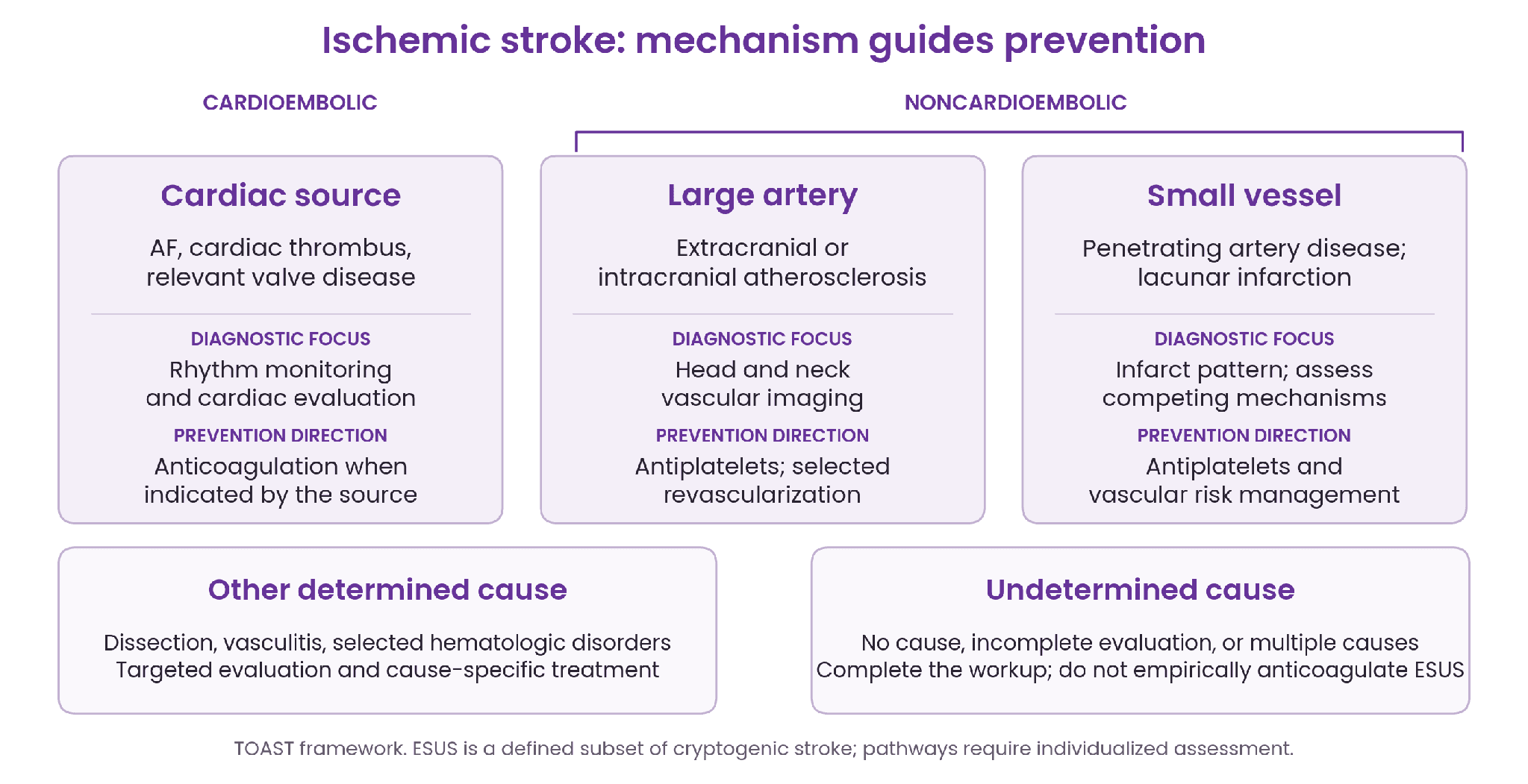
- Cardioembolic: Look for AF, intracardiac thrombus, relevant valve disease, or another high-risk cardiac source. AF is common, but not every cardiac source has the same antithrombotic treatment.
- Noncardioembolic: Large-artery atherosclerosis and small-vessel/lacunar disease generally lead to antiplatelet-based prevention. Vascular imaging distinguishes extracranial from intracranial disease and identifies potential procedural targets.
- Other determined: Consider dissection, vasculitis, and selected hematologic disorders according to age, imaging, and clinical context. Avoid indiscriminate thrombophilia testing; treatment is cause-specific.
- Undetermined: Cryptogenic stroke has no identified cause after evaluation. ESUS is a defined subset with a nonlacunar infarct and no major-risk cardiac source or relevant ≥50% arterial stenosis after a minimum diagnostic workup; it is not synonymous with all cryptogenic stroke.
For hemorrhagic stroke, distinguish intracerebral hemorrhage (ICH) from SAH. Investigate hypertensive arteriopathy, cerebral amyloid angiopathy, antithrombotic exposure, vascular malformations, or aneurysm as appropriate.
Resources & References
Clinical tools and patient education
Guidelines and supporting sources
There’s a place for
your voice here.
Patients and caregivers bring their questions and lived experiences. You bring a perspective that can make a difference. Meet the people behind the questions—and the peers who share your work.